Showing posts with label lumbricals. Show all posts

Saturday, November 26, 2011

Celebrating Advent & Christmas: Trumpet-Organ Duos

Stefan Fleißner
W   hile rotary valve trumpets are not strictly speaking historical but rather contemporary brass instruments, trumpet players have in recent years increasingly been using these instruments for all of the Austro-German classical and romantic repertoire from Mozart and Haydn to Bruckner and Mahler... While the use of rotary valve instruments is certainly not historically accurate for the classical and early romantic natural trumpet orchestral literature, many conductors and players seem to feel that the tapered attack and warmer tone quality of these instruments is better suited to the modern performance of natural trumpet parts than the more brilliant and incisive piston valve instrument. There is no question, however, that the rotary valve trumpet is the stylistically appropriate and historically authentic instrument for the late 19th and early 20th century Germanic repertory.”
  — David H. Green.
T he Advent program at Annakirche in Vienna last night was excellent.

S tefan Fleißner (trpt) and Reinhard Schobesberger (organ) performed a 1-hour program for a delighted audience that completely filled the pews. The trumpet-organ duo works included arrangements of a number of Marienlieder, plus a variety of contemporary Christmas carols, plus the following:
  • Clarke – Trumpet Fanfare, Prinz von Dänemark Marsch
  • Haydn – Andante, Trumpet Concerto in E-flat major
  • Marcello – Concerto in D minor
  • Schubert – Ave Maria
  • Telemann – Trumpet Concerto in D major
  • Bach – ‘Jesu Joy of Man’s Desiring’, from Cantata ‘Herz und Mund und Tat und Leben’, BWV 147
  • Torelli – Sinfonia in D major
I ’ve been in Wien for 7 days, and have attended the Staatsoper, the Wien Philharmoniker, Wien Kammerorchester, Theater an der Wien opera, and various other music events—and I have yet to see a single piston-valved trumpet. Not one. The only thing that anybody uses over here are rotary-valve instruments. Their timbre is very round, full, and capable of a legato sound like nothing else—not as ‘dark’ as a flugelhorn, though, and not as breathy.

T he rotary-valve keys have a shorter ‘throw’ (about 1 cm) than piston-valved instruments, so fingering of rapid passages may, in general, be a bit faster on these instruments, compared to on a conventional piston-valved trumpet. Other biomechanics advantages are mentioned by trumpet players who prefer these rotary-valve designs, including better symmetry between left-hand/arm and right-hand/arm musculature, in terms of holding the instrument… no brass crook for the right-hand “pinky,” and therefore no big forces on that little finger to get transmitted into the lumbricals of the other fingers of that hand; no tension in the left-hand; etc.

Trumpet T he legato playing of Fleißner was superb throughout. Gorgeous, both in the sacred music and in the secular pieces. Annakirche

Saturday, May 22, 2010

Lumbricals, Some Getting Stronger, Others Discouragingly Staying the Same

 Lumbrical muscles
G   reat strength is necessary in the fingers, yes, but it comes with playing, if one plays rightly—that is, musically. From the moment one senses that the finger must ‘sing’, it becomes stronger. That is quite a different matter from playing exercises or etudes merely for the sake of strengthening, and saying ‘I must exercise my fingers and make them strong.’ Such playing as this latter sort does not help at all.”
  —  Vladimir Horowitz.
M y guitarist-pianist friend and colleague at work comes and visits with me this week, says that for more than a month he’s been doing exercises to strengthen the lumbrical muscles in his hands. The left hand has been responding, but the right hand—especially the ring finger and pinky—has been staying the same or, paradoxically, might even be getting weaker.

H e worries about focal dystonia. (Jason Solomon of Georgia Guitar Quartet has an excellent article about that here.) He worries about carpal tunnel syndrome (CTS). I am not a neurologist, but I know enough to know when to go and hire one. I ask my friend about whether he has any numbness or difference in sensation among the fingers on the right hand, or between the right and left...

T he fact that my friend is a professional software developer/engineer who spends 70+ hours at a laptop keyboard each week is something the neurologist will need to know, as part of the evaluation—in addition to the heavy hours on guitar and piano. In other words, if what my friend is experiencing is some type of repetitive stress injury (RSI), then characterizing the various types and intensities of repetitive motions will be clinically important.

T he state-of-the-art of hand biomechanics and hand problems of musicians have been a recurring interest for me for some years, so, in response my friend’s immediate situation, I go online and scan the current medical journal literature, to see what, if anything, is new in the last year or two. For his benefit and maybe for your own, I gather some relevant things together in the list of links below.

W here exactly is the ‘carpal tunnel’? The carpal tunnel is the narrow space anatomically between the small carpal bones of the wrist and the ligament called the flexor retinaculum. Here’s how you can find it: Put your left index finger in the center of your right palm, then move the finger about two inches down your palm toward your arm, stopping when your finger approaches the edge of the fleshy part of your hand. Your finger now lies directly over the carpal tunnel. The carpal tunnel is the U-shaped depression with carpal bones below and on either side. The flexor retinaculum ligament stretches over the top of the ‘U’ to make a tunnel-like space. The cross-section of the tunnel is only a centimeter or so, and nine flexor tendons (two to each finger and one to the thumb) have to pass through that little tunnel. The space is so narrow that some of the tendons are bundled on top of each other instead of going side-by-side the way they do outside the tunnel.

B esides tendons, the median nerve also goes through the carpal tunnel. By contrast, the ulnar nerve does not run in the carpal tunnel. The median nerve supplies most of the palm, the thumb, the index finger, the middle finger, and part of the ring finger. The first and second lumbricals (i.e. the two that are most ‘lateral’ on the radial side; index and middle fingers) are innervated by the median nerve. The third and fourth lumbricals (i.e. the most medial two; middle, ring, and little fingers) are innervated by the deep branch of the ulnar nerve. So if what’s going on is actually CTS, then you might expect weakness predominantly in lumbricals and/or interosseous muscles serving the thumb or index finger or middle finger or maybe the middle fingerward side of the ring finger. And you might think ‘ulnar neuropathy’ if the ring finger and/or pinky are predominantly affected.

B ut, gee, knowledge of the neuroanatomy of peripheral nerves in your arm and wrist and hand only gets you part of the way toward figuring out what is going on. In part, this is because of the interconnections elsewhere, including the motor cortex in your brain. Besides clinical evaluation, electrodiagnostic (EDX) tests are usually needed to confirm the diagnosis.

T he lumbrical and interosseous muscles are important in several motions—including flexing and plucking, increasing and diminishing the ‘spread’ of the fingers, and extending/raising the fingers. The lumbricals are used during an ‘upstroke’ when you are writing with a pen or pencil. These are the muscles that make the fingers separate and spread out or, alternately, converge and come together. The lumbrical muscles, with the help of the interosseous muscles, simultaneously flex the metacarpophalangeal (MCP) joints while extending both interphalangeal (IP) joints. In bats and other animals, these muscles are the ones that enable them to spread the wings and grab the air at one instant and flex and draw them in a few tens of milliseconds later and let the air go. If a bat acquired a repetitive stress injury of its lumbricals, on both sides or one side different from the other, it wouldn’t have long to live. Same thing for a seal: you can’t swim and catch fish if your lumbricals are faltering. Serious musicians—people whose livelihood or soul depends on playing—worry about this, as intently as a seal or bat.

T he EDX testing for these conditions is steadily getting more sensitive and more precise. For example, Sheehan and coworkers (link below) studied people referred with suspected carpal tunnel syndrome (CTS) by measuring the ‘second lumbrical-interosseous distal motor latency difference’ (2LI-DML) as well as by other, more standard tests like ‘median-ulnar palmar velocity difference’. The referred cases included 74% who turned out to be CTS. Sheehan suggests that 2LI-DML, which is a more sensitive test than other nerve conduction velocity tests for detecting mild abnormalities, is useful as a screening test for latent CTS on the asymptomatic side.

M otor distal latency (MDL) differences between the median-thenar and ulnar-hypothenar (M-U) muscles and between the median-second lumbrical and ulnar-interossei muscles (2L-INT) have also recently been used to diagnose early or ‘mixed’ cases. After all, there is no law of Nature that says a person can’t have CTS and UNE or CTS and focal dystonia at the same time. In people in whom the conventional nerve conduction tests are so far ‘normal’ despite the symptoms they are having, the neurologist can measure both motor and sensory W-P conduction and in a large percentage of cases this can establish a diagnosis.

U lnar neuropathy at the elbow (UNE) is the second most common compressive neuropathy of the upper limb. Compared to ‘ulnar neuropathy at the elbow’ (UNE), ulnar neuropathy at the wrist (UNW) is rarer and more difficult to localize with routine electrodiagnostic (EDX) tests. In terms of expectation-setting, it is reasonable to anticipate that it may take some time (and multiple visits) to establish an accurate diagnosis and decide on the right treatment plan. In general, these are not things that can be sorted out in a single, quick office visit.

T he important thing—if you are having symptoms like the ones my guitarist friend is having—is to get yourself examined by a neurologist who is experienced in problems of performing artists and who has the equipment and training to perform the newer EDX tests that are available. You can search for practitioners who are diplomates of the American Board of Electrodiagnostic Medicine here. I regret that I don’t know what comparable search resources there may be for consultants having EDX professional certifications in other countries.


Saturday, February 9, 2008

Strengthening Lumbrical Muscles

Lumbrical muscles
S till having tenderness and weakness in my left hand when I’m playing lute. What exercises should I try? Are there any exercises that I should stay away from—ones that could do more harm than good? Is there any exercise equipment that can make the muscles in my hand stronger, but not make the control coarser or less precise? Wrapping the wrist—would that help maybe? What about wrapping the fingers—especially the middle finger and the ring finger—how about that?”
  — Anonymous.
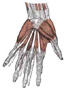
The lumbricals are four small muscles in each hand that interconnect the deep flexor muscles to the extensor muscles of the fingers. The lumbricals are the only muscles in the human body that have no direct bony attachments. They attach at both ends to tendons of these other muscles. The lumbricals are extensors of the proximal interphalangeal (PIP) and distal interphalangeal (DIP) joints. In addition, the lumbricals play a part in side-to-side and rotation movements of the fingers. They’re metacarpal phalangeal (MCP) joint flexors as well, but they’re relatively weak as flexors. They get fatigued easily in their flexor and abductor-adductor and rotator roles.

As each finger moves through its range of motion, each tendon slides a certain distance (An, 1983; Storace and Wolf, 1982). Excursion takes place simultaneously in the flexor and extensor tendons during joint motion: the tendons of the agonist muscles displace in one direction, while the tendons of the antagonist muscles displace in the opposite direction. Knowing what the tendon excursions are has applications for rehabilitation or enhancement of the hand (Brand and Hollister, 1999; Chao, 1989). Most of the time, your exercises should involve motions that cover most of the normal excursion that the tendon (and joint) moves through.


MuscleTendon Excursion (mm)
Interossei 30
Extensor pollicis brevis 30
Abductor pollicis longus 30
Lumbricals 40
Thenar muscles 40
Finger extensors 50-60
Finger flexors 60-70


Coordination is usually tested by counting the number of repetitions of a particular movement—such as rapid repeated tapping—that you can do in 30 seconds. Reduced ability to perform rapid, repetitive movements is often seen for movements that involve muscles like the lumbricals and interossei muscles. Try tapping with each finger of each hand and record your results. The repetitive movement is fatiguing, and you may find you need to concentrate more to perform the test on one side compared with your other side. Lack of precision and lack of smooth movement often are observed, with noticeable differences between fingers and between the two hands. Doing this testing is, admittedly, a bit tedious. But you’ll have a clearer and objective idea of your situation if you do these timed counts.

Subtle issues of coordination and endurance may be more of a problem in lute players than many other musicians—because of the geometry of the instrument. Correcting muscle imbalance requires stretching tight muscles—soft tissue massage. Often a particular area is painful, and ‘trigger points’ are found in the corresponding muscles. Common trigger point sites often include the dorsal first, second, and fourth interossei, the middle finger common extensor, the extensor indicis, the flexor carpi radialis, and flexor carpi ulnaris muscle bellies. You ‘find’ the trigger points by palpating with your right hand’s fingers while you make the left hand do the motions/excursions that are problematic.

Correcting muscle imbalance also requires strengthening the weaker muscles—extensor carpi ulnaris, common wrist extensors, and intrinsic muscles of the hand including the lumbricals. Specific lumbrical muscles surrounding a painful finger are often weak. If you have a lax, hyperextended thumb, it may have a weak extensor pollicis brevis. Often the thumb opponens and flexor pollicis brevis muscles are weak but the thumb adductor muscle is pretty strong. Treatment for hyperlaxity includes temporary supports and strengthening exercises.

Temporary supports to maintain a joint in a neutral position can be helpful. You can gradually wean yourself from them as your symptoms subside and the finger strength improves. Supports may include neoprene wraps or Lycra finger sleeves. These provide joint stability for hypermobile thumb, finger, or wrist joints. The tough thing is to find a support that’s not too bulky or stiff. The reinforced Lycra sleeves that are used for volleyball and other sports aren’t flexible enough. Some Lycra sleeves of the types that are sold in pharmacies are too flexible and don’t offer enough support. Other finger sleeves of the types sold by fly-fishing retailers have a millimeter or two of neoprene or other materials that are too bulky.

DigiSleeve™
Lycra finger sleeves
Stripee™ fly-fishing finger support
The DigiSleeve product is, for me, a reasonable compromise between support and flexibility. It comes as a length of Lycra tube that you cut with a scissors to the exact length that’s right for the dimension of your finger and the amount of the phalanges that you want to cover or support. It’s fairly inexpensive—you just discard the tube segments after several episodes of use and cut yourself new ones. I made a ‘template’ measuring guide on a postcard where I marked off the lengths that I need for the fingers I use the DigiSleeves on, and I keep this template card on my desk next to the box where I keep the DigiSleeve materal. When I need a new sleeve, I just lay the DigiSleeve material on the appropriate template “finger” on the card and cut with a scissors to the right length for that finger. Quick, easy, no re-measuring or guessing, and no wasted material.

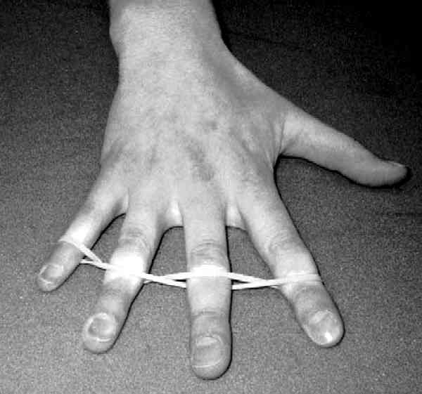
In general, it takes some months for joint stability and lumbrical muscle strength to improve significantly, so you’ll need some patience with the temporary splints or wraps and a modified playing schedule. ‘Stability strengthening’ exercises encourage co-contraction of the muscles surrounding a joint. Improved stability strength helps to compensate for a hyperlax joint. Stability strength training can include isometric and proprioception exercises on your instrument and on exercise equipment like the kinds shown here. For exercises away from the instrument, you can do muscle contractions with a support (such as Lycra finger sleeves) on.

DocZac™ exerciser
GripMaster™ extra-light tension exerciser
In later phases, your hand exercises can include strength training motions that are are concentric and eccentric (with respect to the center of rotation around the affected joint). The DocZac device works pretty well for this. ‘Proprioception’ exercises can include tapping and weightbearing—mostly in a mid-range, neutral joint position. Finger push-ups for hyperlax finger joints, for example—can be performed anytime, against a table or other convenient surface.

x
x
Warrington emphasizes that even the most conscientiously developed exercise program cannot reproduce exactly the action required from the various joints and muscles to play your instrument. And, yes, performing exercises that reproduce some of the actions and that aim to strengthen certain muscles, like the lumbricals, or aim to reduce the laxity of certain joints can be carried too far—excessive exercise can lead to further imbalance or overuse injuries or inflammation, can lead to different problems than the ones the exercises were designed to solve. You’ll be best able to judge your progress—and keep an eye out for problems that may develop—if you keep a log and record your daily hand exercise routine and other relevant details.

Stamina 2.5 output, with left_digit4.txt external input script file
There are fancy, expensive equipments that neurologists use to measure these things. But there are also some freeware/shareware programs that you can use on your computer to do pretty much the same measurements. One of these is Stamina 2.5 (Windows XP / Vista). You can create your own ASCII script files, tailoring the sequence of key-depressions to just those motions that you want to work on or measure, in each of the fingers you are having problems with. From the Stamina 2.5 pull-down Mode menu, choose Other Modes. Select the External File cascading menu choice and navigate to the .txt file you want. Here is one I created for the left ring-finger, and here is one I created for the right ring-finger. Download them and save them to a directory on your PC as you prefer, or to the c:\Program Files\Stamina\Data\ subdirectory where the script files for Stamina 2.5 reside. You can use these as-is, or, more likely, just examine them and see how you may want to set up your own custom script files. Besides revealing the characters-per-minute (cpm) count, which you can record in your daily log, Stamina 2.5 shows the graphical time-trend of how your fingers fatigue with prolonged action. As your stability-strengthening exercises proceed, you may notice that the rate at which your lumbricals-propelled fingers fatigue gets better as the muscles get stronger. And if you experience an injury, the graph is a sensitive indicator of the injury and its severity. The lower cadence achieved by the ring finger may be due more to a lack of independence at the level of voluntary neuromuscular control, than to any innate mechanical interaction with (‘enslavement’ to) the other fingers. Tapping cadence of each finger is lower for the double-finger mode than for the single-finger mode.

You can know for certain when your fingers are being moved by the lumbricals—just make your fingers do a harp-plucking motion. Generally, the lumbricals, flexors, and extensors will all be involved at once, in a way not under your conscious control. Change the finger movement—change the relative amount of straightening and forward motion—and feel your left forearm with your right hand. If the flexor and/or extensor muscles in your arm are contracting, you distinguish it easily because those muscles grow thicker and slide under the skin. When you’re moving your finger forward in such a way that you can’t feel much muscular action in the forearm, then the lumbrical muscle is doing almost all the work. Here are four basic rules for achieving accurate control and endurance:
  1. Do not make unnecessary movements or muscle contractions.
  2. Do the necessary movements as smoothly as possible.
  3. Use the strongest muscles that can do the job.
  4. Exercise to strengthen the muscles that have the potential to do the job.
Good luck!

Tubiana et al.


Sunday, April 29, 2007

Lutes and Lumbricals

Lumbrical Muscles
DSM: A lute player friend of mine has been having progressively more difficulty with his left hand. Focal Dystonia, probably. Nothing so bad as Leon Fleisher’s difficulties years ago or anything like that. But still a major impediment to his performance practice and concertizing.

CMT: There’s still the problem of diagnostic accuracy for Focal Dystonia—and the imprecision of knowing how a loss of “representational specificity” or other changes in the brain can come about through over-use of the hand; the uncertainty of explaining why Botox injected peripherally actually brings about an improvement in this condition if the cause is ‘central’, in the brain. Is your friend sure that it’s Focal Dystonia and not Carpal Tunnel Syndrome or something else?

DSM: Well, no. The neurologists he’s seen have been inconclusive. One hand surgeon wanted to persuade him to be operated for Carpal Tunnel, but my friend regards that as a last resort. And the neurologists’ EMG and imaging and other studies have not been terribly helpful in ruling anything in or out yet. His main complaint has to do with a loss of strength in the lumbrical muscles in his hand, especially the ones involved in moving the middle finger and the ring finger. So it’s not a textbook picture of Focal Dystonia. I suspect he’ll have to go to one of the Performing Arts Medicine clinics that have neurologists experienced in this, as opposed to the well-meaning neurologists and electromyographers he’s seen so far, who have no particular expertise in the problems of musicians.

CMT: There are probably some string players with variant anatomy who just happen to have asymmetrically strong mechanical connections between the ring and middle extrinsic flexor muscles.

Supposedly the phenomenon of middle-ring ‘enslaving’ or ‘subordination’ has a big neural component. If it’s ‘central’ or ‘neural’ then in principle it should be susceptible to retraining through exercises. The connections between adjacent digits involving tendons and muscle fiber groupings of the deep flexor (flexor digitorum profundus) play a big part, though. The ‘peripheral’ biomechanical component shouldn’t be underestimated—it definitely contributes to some of the ‘enslaving’ or ‘subordination’ of one finger to the other. But recent work is showing that enslaving effects among different hand muscles involve both central neural interaction among the structures controlling flexor muscles and lumbricals in the hand as well as peripheral mechanical interactions.

It’s possible that your friend is someone who just happens to have anatomy with substantial tendon sharing. In medical school, I dissected a hand with two lumbrical muscles at each place where you’d ordinarily find only one, and the muscles inserted to adjacent digits. This anatomical variation is not at all uncommon. And if you’re born with this anatomical variation and choose to play lute, then the motions involved in virtuosic lute playing may place particularly great stresses on your aberrant anatomy, in ways that might not affect persons with conventional anatomy quite so severely.

DSM: So what’s going on when we retrain or ‘retune’ the biomechanics with Constraint-Induced Therapy or other physical therapy techniques?

CMT: Well, we learn independent ring and middle finger motion by altering how we activate the controlling networks in the brain, in the primary motor cortex. Neurons in motor cortex aren’t like house-wiring: they don’t make simple direct connections to individual muscles. Instead, our evolution as mammals has set us up to flex and extend our fingers together and to grasp things. Independent digit movements outside of the index finger and thumb require complex inter-neuron coordination to get the desired balance in muscle force at each joint.

Completely independent digit motion is the result of a complex pattern of activation that isolates a movement by preventing other movements mechanically and perhaps by inhibiting some muscles as well. This is why it takes lots of repetitions to learn such skills. Learning new motor skills changes the way muscles and movements are represented topographically among the cells of the motor cortex, and also the way sensory information is represented in the sensory cortex.

Here’s some anatomy, just to give you an idea of where things are, and why the geometry and biomechanics of the hand are so vulnerable as they are.

Crossection of hand, 4 fingers, distal to thumb


Upper Left

Lower Left

Lower Right

Upper Right


1. Distal palmar fat pad

2. First lumbrical m.

3. Tendons mm. flexors digitorum superficialis and profundus

4. Tendons mm. interosseous
and lumbrical


5. Proximal phalanx, second digit

6. Second lumbrical m.


7. Tendon extensor digitorum communis

8. Dorsal and palmar interosseous mm.

9. Tendon m. extensor indicis proprius

10. Collateral ligament

11. Tendons mm. interosseous
and lumbrical


12. Proximal phalanx, third digit

13. Tendon m. extensor digitorum communis


14. Tendons and mm. dorsal
and palmar interosseous

15. Tendons mm. interosseous
and lumbrical


16. Tendon m. extensor digitorum communis

17. Proximal phalanx, fourth digit

18. Tendons mm. interosseous
and lumbrical


19. Tendons mm. flexors digitorum
superficialis and profundus


20. Proximal phalanx, fifth digit

21. Articular capsule and collateral ligament

22. Interphalangeal joint

23. Middle phalanx, fifth digit

24. Fifth digit, distal segment fat pad

25. Fibrous digital sheath

26. Tendon m. flexor digitorum
profundus


The photomacrograph of a transverse whole-section of the hand passes through the four fingers. The slice is taken of a cadaver hand, a frozen section, distal to the thumb—right at the end of the hand, before the fingers start. You can see that the little finger is separated from the others, which makes sense if you look at your own hand. The gap between the little finger and the ring finger comes proximally further into the hand, compared to the gaps between the other fingers. The articulation (22) between the proximal (20) and middle (23) phalanges is seen in the cross-sectional slice in the picture above. This is a pretty bulky hand. Probably not the hand of a musician. But the image helps to give you and idea of the anatomical relationships just the same.

Notice how the extensor tendon to the ring (fourth) finger is very closely applied to its dorsal surface. Not much clearance there! The tendons and distal ends of both interosseous and lumbrical muscles (8, 11, 14, 15, 18) are identified in the picture in relation to the middle digit proximal phalanx (12). The first (2) and second (6) lumbricals are seen close to, and on the radial side of, their tendons of origin. The two lateral (first and second) lumbricals arise from the radial side of the palmer aspect of the first and second tendons of flexor digitorum profundus. The two medial (third and fourth) arise from adjacent sides of the second and third, and the third and fourth tendons of flexor digitorum profundus. The muscles insert into the radial border of the tendon of extensor digitorum on the back of the proximal phalanx. The compactness of this anatomy reveals why the mechanical leverage and pulley-and-lever structures are so exquisitely sensitive. The fulcrums for the action of the lumbricals on the phalanges operate at a relatively high mechanical advantage (force ratio), and therefore small disturbances in the muscles themselves or in the tendon bundle and the tendons’ insertion points can give rise to surprisingly large disturbances in the motion—velocity of attack and release, power, etc.—at the phalanx.

Principles of Neuroplasticity—Strategies for Rehabilitation for Focal Dystonia through Forced Use:
  • Based on the assumption that patients will more readily use the extremity that is normal (unaffected) unless you restrict it.
  • Forcing the use of the affected extremity will modify muscle structure and increase function.
  • Effective for pianists but not for guitarists or lutenists.

Constraint-Induced Therapy (Sensory Motor Retuning):
  • Elbert, Sterr, and colleagues studied 7 musicians with Focal Dystonia.
  • Forced use on the instrument with a splint that restricted the movement of each finger; 10 days, 7 hrs each day.
  • Six of 7 improved; less improvement for guitarists or lutenists than for pianists.

In the absence of a definitive diagnosis, the Physical Therapist or Rehabilitation Medicine practitioner may treat on an empirical basis:
  • Treat the initial injury as part of [an empirically conjectured—] inflammatory, healing process.
  • Decrease stress and stop negative abnormal patterns of movement.

At least in me, there are strong linkages between the middle and ring fingers when I attempt to flex the middle finger at the distal (end) joints. This probably involves mechanical linkages between the profundus muscles for those two fingers—linkages at the level of both proximal tendon and muscle. Nobody knows whether those linkages can evolve over time under conditions of high repetition stress or over-use.

DSM: Obviously, if inflammation is present, you’d expect that some fibrosis and linkage would occur as part of the healing and response to the inflammation.

CMT: But if no overt tendonitis or other inflammatory process is going on, it’s not clear whether a fibrotic proliferative reaction would occur, to give rise to the kind of symptoms your friend is experiencing. Assuming it’s not Focal Dystonia, the Constraint-Induced Therapy of “retuning” exercises can help to compensate for any abnormal anatomical linkages that might be in his hand. Best of luck to your lutenist friend!